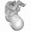
Prenatal development is divided into two primary biological stages. The first is the embryonic stage, which lasts for about two months. At this point, the fetal stage begins. At the beginning of the fetal stage, the risk of miscarriage decreases sharply,[36] all major structures including the head, brain, hands, feet, and other organs are present, and they continue to grow and develop. When the fetal stage commences, a fetus is typically about 30 mm (1.2 inches) in length, and the heart can be seen beating via sonograph; the fetus bends the head, and also makes general movements and startles that involve the whole body.[37] Some fingerprint formation occurs from the beginning of the fetal stage.[38]
Electrical brain activity is first detected between the 5th and 6th week of gestation, though this is still considered primitive neural activity rather than the beginning of conscious thought, something that develops much later in fetation. Synapses begin forming at 17 weeks, and at about week 28 begin to multiply at a rapid pace which continues until 3–4 months after birth. It is not until week 23 that the fetus can survive, albeit with major medical support, outside of the womb, because it does not possess a sustainable human brain until that time.[39]
One way to observe prenatal development is via ultrasound images. Modern 3D ultrasound images provide greater detail for prenatal diagnosis than the older 2D ultrasound technology.[44] While 3D is popular with parents desiring a prenatal photograph as a keepsake,[45] both 2D and 3D are discouraged by the FDA for non-medical use,[46][dead link] but there are no definitive studies linking ultrasound to any adverse medical effects.[47] The following 3D ultrasound images were taken at different stages of pregnancy:
- 75-mm fetus (about 14 weeks gestational age)
Some people are confused about the differences between an ultrasound and a sonogram. An ultrasound is the actual machine that lets you observe pregnancy. A sonogram is the image of the baby that the ultrasound produces. 4D Ultrasounds take 3D sonograms. Some people refer to the procedure as prenatal imaging, 3D imaging, a 3D scan, or 4D scan.
 1:23 AM
 kotesh


