BIOTECHNOLOGY


Biotechnology is a field of applied biology that involves the use of living organisms and bioprocesses in engineeringtechnologymedicine and other fields requiring bioproducts.Biotechnology also utilizes these products for manufacturing purpose. Modern use of similar terms includes genetic engineering as well as cell- and tissue culture technologies. The concept encompasses a wide range of procedures (and history) for modifying living organisms according to human purposes - going back to domestication of animals, cultivation of plants, and "improvements" to these through breeding programs that employ artificial selection and hybridization. By comparison to biotechnology, bioengineering is generally thought of as a related field with its emphasis more on higher systems approaches (not necessarily altering or using biological materials directly) for interfacing with and utilizing living things. The United Nations Convention on Biological Diversity defines biotechnology as:[1]
"Any technological application that uses biological systems, living organisms, or derivatives thereof, to make or modify products or processes for specific use."
In other term "Application of scientific and technical advances in life science to develop commercial products" is biotechnology.
Biotechnology draws on the pure biological sciences (geneticsmicrobiologyanimal cell culturemolecular biologybiochemistryembryologycell biology) and in many instances is also dependent on knowledge and methods from outside the sphere of biology (chemical engineeringbioprocess engineeringinformation technologybiorobotics). Conversely, modern biological sciences (including even concepts such as molecular ecology) are intimately entwined and dependent on the methods developed through biotechnology and what is commonly thought of as the life sciences industry.

Sexual activity during pregnancy

Most pregnant women can enjoy sexual activity during pregnancy throughout gravidity. Most research suggests that, during pregnancy, both sexual desire and frequency of sexual relations decrease.[75][76] In context of this overall decrease in desire, some studies indicate a second-trimester increase, preceding a decrease.[77][78] However, these decreases are not universal: a significant number of women report greater sexual satisfaction throughout their pregnancies.

medicine which used during Pregnancy

Drugs used during pregnancy can have temporary or permanent effects on the fetus. Therefore many physicians would prefer not to prescribe for pregnant women, the major concern being over teratogenicity of the drugs.
Drugs have been classified into categories A,B,C,D and X based on the Food and Drug Administration (FDA) rating system to provide therapeutic guidance based on potential benefits and fetal risks. Drugs like multivitaminsthalidomide with proven fetal risks that outweigh all benefits are classified as Category X that have demonstrated no fetal risks after controlled studies in humans are classified as Category A. On the other hand drugs like

Physiological changes of Pregnancy

During pregnancy, the woman undergoes many physiological changes, which are entirely normal, including cardiovascular, hematologic, metabolic, renal and respiratory changes that become very important in the event of complications. The body must change its physiological and homeostatic mechanisms in pregnancy to ensure the fetus is provided for. Increases in blood sugar, breathing and cardiac output are all required. Levels of progesterone and oestrogens rise continually throughout pregnancy, suppressing the hypothalamic axis and subsequently the menstrual cycle. The woman and the placenta also produce many hormones.

Embryonic and fetal development of Pregnancy

Prenatal development is divided into two primary biological stages. The first is the embryonic stage, which lasts for about two months. At this point, the fetal stage begins. At the beginning of the fetal stage, the risk of miscarriage decreases sharply,[36] all major structures including the head, brain, hands, feet, and other organs are present, and they continue to grow and develop. When the fetal stage commences, a fetus is typically about 30 mm (1.2 inches) in length, and the heart can be seen beating via sonograph; the fetus bends the head, and also makes general movements and startles that involve the whole body.[37] Some fingerprint formation occurs from the beginning of the fetal stage.[38]
Electrical brain activity is first detected between the 5th and 6th week of gestation, though this is still considered primitive neural activity rather than the beginning of conscious thought, something that develops much later in fetation. Synapses begin forming at 17 weeks, and at about week 28 begin to multiply at a rapid pace which continues until 3–4 months after birth. It is not until week 23 that the fetus can survive, albeit with major medical support, outside of the womb, because it does not possess a sustainable human brain until that time.[39]
One way to observe prenatal development is via ultrasound images. Modern 3D ultrasound images provide greater detail for prenatal diagnosis than the older 2D ultrasound technology.[44] While 3D is popular with parents desiring a prenatal photograph as a keepsake,[45] both 2D and 3D are discouraged by the FDA for non-medical use,[46][dead link] but there are no definitive studies linking ultrasound to any adverse medical effects.[47] The following 3D ultrasound images were taken at different stages of pregnancy:
Some people are confused about the differences between an ultrasound and a sonogram. An ultrasound is the actual machine that lets you observe pregnancy. A sonogram is the image of the baby that the ultrasound produces. 4D Ultrasounds take 3D sonograms. Some people refer to the procedure as prenatal imaging, 3D imaging, a 3D scan, or 4D scan.

Third Trimester of Pregnancy

Final weight gain takes place, which is the most weight gain throughout the pregnancy. The fetus will be growing the most rapidly during this stage, gaining up to 28 g per day. The woman's belly will transform in shape as the belly drops due to the fetus turning in a downward position ready for birth. During the second trimester, the woman's belly would have been very upright, whereas in the third trimester it will drop down quite low, and the woman will be able to lift her belly up and down. The fetus begins to move regularly, and is felt by the woman. Fetal movement can become quite strong and be disruptive to the woman. The woman's navel will sometimes become convex, "popping" out, due to her expanding abdomen. This period of her pregnancy can be uncomfortable, causing symptoms like weak bladder control and backache. Movement of the fetus becomes stronger and more frequent and via improved brain, eye, and muscle function the fetus is prepared for ex utero viability. The woman can feel the fetus "rolling" and it may cause pain or discomfort when it is near the woman's ribs and spine.
 
There is head engagement in the third trimester, that is, the fetal head descends into the pelvic cavity so that only a small part (or none) of it can be felt abdominally. The perenium and cervix are further flattened and the head may be felt vaginally.[33] Head engagement is known colloquially as the baby drop, and in natural medicine as the lightening because of the release of pressure on the upper abdomen and renewed ease in breathing. However, it severely reduces bladder capacity, increases pressure on the pelvic floor and the rectum, and the mother may experience the perpetual sensation that the fetus will "fall out" at any moment.[34]
It is during this time that a baby born prematurely may survive. The use of modern medical intensive care[35] In spite of these developments, premature birth remains a major threat to the fetus, and may result in ill health in later life, even if the baby survives. technology has greatly increased the probability of premature babies surviving, and has pushed back the boundary of viability to much earlier dates than would be possible without assistance.

Second Trimester of Pregnancy

Weeks 13 to 28 of the pregnancy are called the second trimester. Most women feel more energized in this period, and begin to put on weight as the symptoms of morning sickness subside and eventually fade away.
In the 20th week, the uterus, the muscular organ that holds the developing fetus, can expand up to 20 times its normal size during pregnancy. Although the fetus begins to move and takes a recognizable human shape during the first trimester, it is not until the second trimester that movement of the fetus, often referred to as "quickening", can be felt. This typically happens in the fourth month, more specifically in the 20th to 21st week, or by the 19th week if the woman has been pregnant before. However, it is not uncommon for some women not to feel the fetus move until much later. The placenta fully functions at this time and the fetus makes insulin and urinates. The reproductive organs distinguish the fetus as male or female.

First trimester of Pregnancy

First trimester

Traditionally, doctors have measured pregnancy from a number of convenient points, including the day of last menstruation, ovulation, fertilization, implantation and chemical detection. In medicine, pregnancy is often defined as beginning when the developing embryo becomes implanted into the endometrial lining of a woman's uterus. In some cases where complications may have arisen, the fertilized egg might implant itself in the fallopian tubes, the cervix, the ovary or in the abdomen causing an ectopic pregnancy. In the case of an ectopic pregnancy there is no way for the pregnancy to progress normally. If left untreated, it can cause harm and possibly death for the mother when a rupture occurs. Sometimes it will go away on its own but otherwise a surgical procedure or medicine is given to remove the tubal pregnancy since there is no way of the pregnancy being able to continue safely.[28] Most pregnant women do not have any specific signs or symptoms of implantation, although it is not uncommon to experience minimal bleeding at implantation. Some women will also experience cramping during their first trimester. This is usually of no concern unless there is spotting or bleeding as well. After implantation the uterine endometrium is called the decidua. The placenta, which is formed partly from the decidua and partly from outer layers of the embryo, connects the developing fetus to the uterine wall to allow nutrient uptake, waste elimination, and gas exchange via the mother's blood supply. The umbilical cord is the connecting cord from the embryo or fetus to the placenta. The developing embryo undergoes tremendous growth and changes during the process of fetal development.
Morning sickness occurs in about seventy percent of all pregnant women and typically improves after the first trimester.[29][dead link] Although described as "morning sickness", women can experience this nausea during afternoon, evening, and throughout the entire day.
In the first 12 weeks of pregnancy, the nipples and areolas darken due to a temporary increase in hormones.[30]
The first 12 weeks of pregnancy are considered to make up the first trimester. The first two weeks from the first trimester are calculated as the first two weeks of pregnancy even though the pregnancy does not actually exist. These two weeks are the two weeks before conception and include the woman's last period.
The third week is the week in which fertilization occurs and the 4th week is the period when implantationuterus and burrows into its wall which provides it with the nutrients it needs. At this point, the zygote becomes a blastocyst and the placenta starts to form. Moreover, most of the pregnancy tests may detect a pregnancy beginning with this week. takes place. In the 4th week, the fecundated egg reaches the
The 5th week marks the start of the embryonic period. This is when the baby's brain, spinal cord, heart and other organs begin to form.[31] At this point the embryo is made up of three layers, of which the top one (called the ectoderm) will give rise to the baby's outermost layer of skin, central and peripheral nervous systems, eyes, inner ear, and many connective tissues.[31] The heart and the beginning of the circulatory system as well as the bones, muscles and kidneys are made up from the mesoderm (the middle layer). The inner layer of the embryo will serve as the starting point for the development of the baby's lungs, intestine and bladder. This layer is referred to as the endoderm. A baby at 5 weeks is normally between 116 and 18 inch (1.6 and 3.2 mm) in length.
In the 6th week, the baby will be developing basic facial features and its arms and legs start to grow. At this point, the embryo is usually no longer than 16 to 14 inch (4.2 to 6.3 mm). In the following week, the brain, face and arms and legs quickly develop. In the 8th week, the baby starts moving and in the next 3 weeks, the baby's toes, neck and genitals develop as well. According to the American Pregnancy Association, by the end of the first trimester, the fetus will be about 3 inches (76 mm) long and will weigh approximately 1 ounce (28 g).[32]

Child Birth

Childbirth is the process whereby an infant is born. It is considered by many[who?] to be the beginning of the infant's life, and age is defined relative to this event in most cultures.
A woman is considered to be in labour when she begins experiencing regular uterine contractions, accompanied by changes of her cervix — primarily effacement and dilation. While childbirth is widely experienced as painful, some women do report painless labours, while others find that concentrating on the birth helps to quicken labour and lessen the sensations. Most births are successful vaginal births, but sometimes complications arise and a woman may undergo a cesarean section.
During the time immediately after birth, both the mother and the baby are hormonally cued to bond, the mother through the release of oxytocin, a hormone also released during breastfeeding.

Duration of Pregnancy


The expected date of delivery (EDD) is 40 weeks counting from the first day of the last menstrual period[13] The actual pregnancy duration is typically 38 weeks after conception. Though pregnancy begins at conception, it is more convenient to date from the first day of a woman's last menstrual period, or from the date of conception if known. Starting from one of these dates, the expected date of delivery can be calculated using the Naegele's rule for estimating date of delivery. A more accurate and sophisticated algorithm takes into account other variables, such as whether this is the first or subsequent child (i.e., pregnant woman is a primip or a multip, respectively), ethnicity, parental age, length of menstrual cycle, and menstrual regularity. (LMP), and birth usually occurs between 37 and 42 weeks.
Pregnancy is considered "at term" when gestation attains 37 complete weeks but is less than 42 (between 259 and 294 days since LMP). Events before completion of 37 weeks (259 days) are considered preterm; from week 42 (294 days) events are considered postterm.[14] When a pregnancy exceeds 42 weeks (294 days), the risk of complications for both the woman and the fetus increases significantly.[13][15] As such, obstetricians usually prefer to induce labour, in an uncomplicated pregnancy, at some stage between 41 and 42 weeks.[16][17]
Recent medical literature prefers the terminology preterm and postterm to premature and postmature. preterm and postterm are unambiguously defined as above, whereas premature and postmature have historical meaning and relate more to the infant's size and state of development rather than to the stage of pregnancy.[18][19]
Fewer than 5% of births occur on the due date; 50% of births are within a week of the due date, and almost 90% within 2 weeks.[unreliable medical source?][20] It is much more useful and accurate, therefore, to consider a range of due dates, rather than one specific day, with some online due date calculators providing this information.
Accurate dating of pregnancy is important, because it is used in calculating the results of various prenatal teststriple test). A decision may be made to induce labour if a fetus is perceived to be overdue. Furthermore, if LMP and ultrasound dating predict different respective due dates, with the latter being later, this might signify slowed fetal growth and therefore require closer review. (for example, in the
The age of viability has been receding because of continued medical progress. Whereas it used to be 28 weeks, it has been brought back to as early as 23, or even 22 weeks in some countries.

prenetal Period

Prenatal defines the period occurring "around the time of birth", specifically from 22 completed weeks (154 days) of gestation (the time when birth weight is normally 500 g) to 7 completed days after birth.[12]
Legal regulations in different countries include gestation age beginning from 16 to 22 weeks (5 months) before birth.
The perinatal period is immediately before to after birth. Depending on the definition, it starts between the 20th to 28th week of gestation and ends between 1 to 4 weeks after birth (the word "perinatal" is a hybrid of the Greek "peri-" meaning 'around or about' and "natal" from the Latin "natus" meaning "birth.").

What is Pregnancy ?

Pregnancy is the carrying of one or more offspring, known as a fetus or embryo, inside the womb of a female. In a pregnancy, there can be multiple gestations, as in the case of twins or triplets. Human pregnancy is the most studied of all mammalian pregnancies. Childbirth usually occurs about 38 weeks after conception; in women who have a menstrual cycle length of four weeks, this is approximately 40 weeks from the last normal menstrual period (LNMP). The World Health Organization defines normal term for delivery as between 37 weeks and 42 weeks.

A World Without Sex?


The birth of Louise, whose twentieth birthday was celebrated in Britain with great fanfare, excited fears and controversy similar to those surrounding cloning (see Genetic Engineering). As with cloning, the fear was that the technology of in vitro fertilization would lead to the depersonalized manufacturing of human beings associated with some nightmarish future society, but this has not come to pass for several reasons. One is that in vitro fertilization is successful only about 15% of the time. Another, much more significant reason is that there are few women capable of producing a child through internal fertilization who would want to conceive without the intimacy of sexual intercourse.
There are exceptions, of course, in real life (two women in a same-sex relationship who want a child, for example) as well as in fiction. In the latter category, the character of Jenny Fields in the American novelist John Irving's World According to Garp—published, ironically, in the same year as the birth of Louise Brown—would undoubtedly have conceived a child by in vitro means if those means had been available to her. As it was, Jenny, who conceived the book's title character during World War II, manages to do so by having intercourse purely for the purpose of producing offspring. She does this by choosing a wounded, brain-damaged airman who will never remember having had sex with her and who dies shortly thereafter.

External Fertilization


Most land animals use some form of internal fertilization similar to that which we have described for humans. External fertilization, on the other hand, is more common among aquatic animals, who simply dump their sperm and eggs into the water and let currents mix the two male and female cells together. The sea urchin is a typical example: a male sea urchin releases several billion sperm into the water, and these sperm then swim toward eggs released in the same area. Fertilization occurs within seconds when sperm come into contact and fuse with eggs. As noted in Reproduction, external fertilization is essentially sexual reproduction without sexual intercourse. For humans the process of reproduction by external means may lack the intimacy of internal reproduction, but since 1978 a form of external fertilization has offered the opportunity of conceiving children to couples who otherwise might have remained childless.

The Ovaries and Menstruation


Eggs are produced in the ovaries, oval-shaped organs in the groin that also generate sex hormones. At birth, a female's ovaries contain hundreds of thousands of undeveloped eggs, each surrounded by a group of cells to form a follicle, or sac; however, only about 360-480 follicles reach full maturity. During puberty the action of hormones causes several follicles to develop each month. Normally, just one follicle fully matures, rupturing and releasing an ovum through the ovary wall in a process called ovulation. The mature egg enters one of the paired fallopian tubes, where it may be fertilized by a sperm and move on to the uterus to develop into a fetus. The lining of the uterus, called the endometrium, prepares for pregnancy each month by thickening, but if fertilization does not take place, the endometrium is shed during menstruation.

The Reproductive System


The contrast between sexual and asexual reproduction is examined in Reproduction, an essay that also provides examples of plant reproduction through pollination. The present essay is concerned primarily with human sexual reproduction and secondarily with animal sexual reproduction. Some technical aspects of reproduction at the cellular level require consultation of processes explained in Genetics; here we confine our technical discussion to reproduction at the level of organs, fluids, and other bodily components. Reproduction is facilitated by the reproductive system, a group of organized structures that can be subdivided into male and female reproductive systems. During puberty, which typically occurs between the ages of 10 and 14 years, the reproductive systems of both sexes mature. This phase is marked in part by the release of eggs (female sex cells) in the female ovary and the formation of sperm (male sex cells) in the male testes. Reproduction can take place only when a sperm unites with an egg, a process called fertilization.

Why Sexual Reproduction?

When Maynard Smith reverse-engineered sex... he created a paradox. Sex should not exist; natural selection will favor asexual reproduction. The solution to the paradox is almost the Holy Grail of a large theoretical sub-branch of evolutionary biology, but it still has not been satisfactorily tracked down. — Mark Ridley, 1997 (1)
In spite of the number of ways bacteria can exchange genetic instructions, many of them haven't evolved very far — not as bacteria anyway. There are many modern bacteria that look very similar to fossils of the earliest bacteria of over 3 billion years ago. Eukaryotes are much better at evolving than prokaryotes. Eukaryotes first appeared on Earth about 1.7 billion years ago. They contain complex subunits, some of which — mitochondria and plastids — have their own DNA. With the appearance of eukaryotes, evolution began to accelerate. All multicelled animals and plants are made of eukaryotic cells. Where did they come from?

SEXUAL REPRODUCTION IN FISH

The vast majority of fish species lay eggs that are then fertilized by the male,[6] some species lay their eggs on a substrate like a rock or on plants, while others scatter their eggs and the eggs are fertilized as they drift or sink in the water column. Some fish species use internal fertilization and then disperse the developing eggs or give birth to live offspring. Fish that have live-bearing offspring include the Guppy and Mollies or Poecilia. Fishes that give birth to live young can be ovoviviparous, where the eggs are fertilized within the female and the eggs simply hatch within the female body, or in seahorses, the male carries the developing young within a pouch, and gives birth to live young.[7] Fishes can also be viviparous, where the female supplies nourishment to the internally growing offspring. Some fish are hermaphrodites, where a single fish is both male and female and can produce eggs and sperm. In hermaphroditic fish, some are male and female at the same time while in other fish they are serially hermaphroditic; starting as one sex and changing to the other. In at least one hermaphroditic species, self-fertilization occurs when the eggs and sperm are released together. Internal self-fertilization may occur in some other species.[8] One fish species does not reproduce by sexual reproduction but uses sex to produce offspring; Poecilia formosa is a unisex species that uses a form of parthenogenesis called gynogenesis, where unfertilized eggs develop into embryos that produce female offspring. Poecilia formosa mate with males of other fish species that use internal fertilization, the sperm does not fertilize the eggs but stimulates the growth of the eggs which develops into embryos.[9]

FEMALE REPRODUCTIVE SYSTEM

The female reproductive system likewise contains two main divisions: the vagina and uterus, which act as the receptacle for the sperm, and the ovaries, which produce the female's ova. All of these parts are always internal. The vagina is attached to the uterus through the cervix, while the uterus is attached to the ovaries via the Fallopian tubes. At certain intervals, the ovaries release an ovum, which passes through the fallopian tube into the uterus.
If, in this transit, it meets with sperm, the egg selects sperm with which to merge; this is termed fertilization. The fertilization usually occurs in the oviducts, but can happen in the uterus itself. The zygote then implantsembryogenesis and morphogenesis. When developed enough to survive outside the womb, the cervix dilates and contractions of the uterus propel the fetus through the birth canal, which is the vagina. itself in the wall of the uterus, where it begins the processes of
The ova, which are the female sex cells, are much larger than the sperm and are normally formed within the ovaries of the fetus before its birth. They are mostly fixed in location within the ovary until their transit to the uterus, and contain nutrients for the later zygote and embryo. Over a regular interval, in response to hormonal signals, a process of oogenesis matures one ovum which is released and sent down the Fallopian tube. If not fertilized, this egg is released through menstruation in humans and other great apes, and reabsorbed in other mammals in the estrus cycle.

MALE REPRODUCTIVE SYSTEM

The male reproductive system contains two main divisions: the penis, and the testicles, the latter of which is where sperm are produced. In humans, both of these organs are outside the abdominal cavity, but they can be primarily housed within the abdomen in other animals (for instance, in dogs, the penis is internal except when mating). Having the testicles outside the abdomen best facilitates temperature regulation of the sperm, which require specific temperatures to survive. Sperm are the smaller of the two gametes and are generally very short-lived, requiring males to produce them continuously from the time of sexual maturity until death. Prior to ejaculation the produced sperm are stored in the epididymis. The sperm cells are motile and they swim using tail-like flagella to propel themselves towards the ovum. The sperm follows temperature gradients (thermotaxis)[3] and chemical gradients (chemotaxis) to locate the ovum.

SEXUAL REPRODUCTION IN INSECTS

Insect species make up more than two-thirds of all extant animal species, and most insect species use sex for reproduction, though some species are facultatively parthenogenetic. Many species have sexual dimorphism, while in others the sexes look nearly identical. Typically they have two sexes with males producing spermatozoa and females ova. The ova develop into eggs that have a covering called the chorion, which forms before internal fertilization. Insects have very diverse mating and reproductive strategies most often resulting in the male depositing spermatophore within the female, which stores the sperm until she is ready for egg fertilization. After fertilization, and the formation of a zygote, and varying degrees of development; the eggs are deposited outside the female in many species, or in some, they develop further within the female and live born offspring are produced.

SEXUAL REPRODUCTION OF HUMAN BEINGS

Sexual reproduction is the creation of a new organism by combining the genetic material of two organisms. The two main processes are: meiosis, involving the halving of the number of chromosomes; and fertilization, involving the fusion of two gametes and the restoration of the original number of chromosomes. During meiosis, the chromosomes of each pair usually cross over to achieve homologous recombination.
The evolution of sexual reproduction is a major puzzle. The first fossilized evidence of sexually reproducing organisms is from eukaryotes of the Stenian period, about 1 to 1.2 billion years ago.[1] Sexual reproduction is the primary method of reproduction for the vast majority of macroscopic organisms, including almost all animals and plants. Bacterial conjugation, the transfer of DNA between two bacteria, is often mistakenly confused with sexual reproduction, because the mechanics are similar.
A major question is why sexual reproduction persists when parthenogenesis appears in some ways to be a superior form of reproduction. Contemporary evolutionary thought proposes some explanations. It may be due to selection pressure on the clade itself—the ability for a population to radiate more rapidly in response to a changing environment through sexual recombination than parthenogenesis allows. Alternatively, sexual reproduction may allow for the "ratcheting" of evolutionary speed as one clade competes with another for a limited resource.

RADIATION TREATMENT FOR BRAIN TUMOR....!

The goal of radiation therapy is to selectively kill tumor cells while leaving normal brain tissue unharmed. In standard external beam radiation therapy, multiple treatments of standard-dose "fractions" of radiation are applied to the brain. Each treatment induces damage to both healthy and normal tissue. By the time the next treatment is given, most of the normal cells have repaired the damage, but the tumor tissue has not. This process is repeated for a total of 10 to 30 treatments, depending on the type of tumor. This additional treatment provides some patients with improved outcomes and longer survival rates.
Radiosurgery is a treatment method that uses computerized calculations to focus radiation at the site of the tumor while minimizing the radiation dose to the surrounding brain. Radiosurgery may be an adjunct to other treatments, or it may represent the primary treatment technique for some tumors.
Radiotherapy may be used if there is incomplete resection of the tumor. Forms of radiotherapy used for brain cancer include external beam radiation therapy, brachytherapy ,and in more difficult cases, stereotacticradiosurgery, such as Gamma knife, Cyberknife or Novalis Tx radiosurgery, remains a viable option.[15]
Radiotherapy is the most common treatment for secondary cancer brain tumors. The amount of radiotherapy depends on the size of the area of the brain affected by cancer. Conventional external beam whole brain radiotherapy treatment (WBRT) or 'whole brain irradiation' may be suggested if there is a risk that other secondary tumors will develop in the future.[16] Stereotactic radiotherapy is usually recommended in cases of under three small secondary brain tumors.
In 2008 a study published by the University of Texas M. D. Anderson Cancer Center indicated that cancer patients who receive stereotactic radiosurgery (SRS) and whole brain radiation therapy (WBRT) for the treatment of metastatic brain tumors have more than twice the risk of developing learning and memory problems than those treated with SRS alone

SURGERIES FOR BRAIN TUMOR'S

The primary course of action described in medical literature is surgical removal (ressection), a craniotomy(to open the skull) to best access the tumor site, currently minimal invasive techniques are being studied but far from being common practice. The prime re mediating objective of surgery is to remove as many tumor cells as possible, in any case surgery will reduce the tumor size and will attempt to completely remove the neoplasm. In some cases access to the tumor is impossible and impedes surgery.
Many meningiomas, with the exception of some tumors located at the skull base, can be successfully removed surgically. Most can be removed surgically, often using a minimally invasive approach through the nasal cavity and skull base (trans-nasal, trans-sphenoidal approach). Large pituitary adenomascraniotomy (opening of the skull) for their removal. Radiotherapy, including stereotactic approaches, is reserved for the inoperable cases. require a
Although there is no generally accepted therapeutic management for primary brain tumors, a surgical attempt at tumor removal or at least cytoreduction (that is, removal of as much tumor as possible, in order to reduce the number of tumor cells available for proliferation) is considered in most cases.[13] However, due to the infiltrative nature of these lesions, tumor recurrence, even following an apparently complete surgical removal, is not uncommon. Several current research studies aim to improve the surgical removal of brain tumors by labeling tumor cells with a chemical (5-aminolevulinic acid) that causes them to fluoresce.[14] Postoperative radiotherapy and chemotherapy are integral parts of the therapeutic standard for malignant tumors. Radiotherapy may also be administered in cases of "low-grade" gliomas, when a significant tumor burden reduction could not be achieved surgically.
The main treatment option for single metastatic tumors is surgical removal, followed by radiotherapy and/or chemotherapy. Multiple metastatic tumors are generally treated with radiotherapy and chemotherapy. Stereotactic radiosurgery (SRS), such as Gamma Knife, Cyberknife or Novalis Tx, radiosurgery, remains a viable option. However, the prognosis in such cases is determined by the primary tumor, and it is generally poor.

Diagnosis

Although there is no specific or singular clinical symptom or sign for any brain tumors, the presence of a combination of symptoms and the lack of corresponding clinical indications of infections might be an indicator to step up the diagnostic investigation to the direction of an intracranial neoplasm.
The diagnosis will often start with an interrogation of the patient to get a clear view of his medical antecedents, and his current symptoms. Clinical and laboratory investigations will serve to exclude infections as cause of the symptoms. Examinations in this stage may include ophtamological, otolaryngological (or ENT) and/or Electrophysiological exams, other means such as electroencephalography (EEG) play a role in the diagnosis of brain tumors.
Swelling, or obstructing the passage of cerebrospinal fluid (CSF) may cause (early) signs of increased intracranial pressure which translates clinically into headaches, vomiting, or an altered state of consciousness, (and in children) changes to the diameter of the skull and bulging of the fontanelles. More complex symptoms such as endocrine dysfunctions should alarm doctors not to exclude brain tumors.
A bilateral temporal visual field defect (due to compression of the optic chiasm) or dilatation of the pupil, and the occurrence of either slowly evolving or the sudden onset of focal neurologic symptoms, such as cognitivebehavioral impairment (including impaired judgment, memory loss, lack of recognition, spatial orientation disorders), personality or emotional changes, hemiparesis, hypoesthesia, aphasia, ataxia, visual fieldfacial paralysis, double vision, but also more severe symptoms might accur too such as: tremors, paralysis on one side of the body hemiplegia, but also (epileptic) seizures in a patient with a negative history for epilepsy, impairment to swallow should raise red flags. and impairment, impaired sense of smell, impaired hearing,

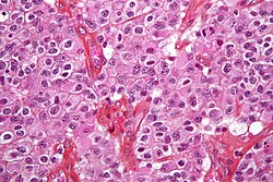
Micrograph of an oligodendroglioma, a type of brain cancer. Brain biopsy. H&E stain.
Imaging plays a central role in the diagnosis of brain tumors. Early imaging methods —invasive and sometimes dangerous— such as pneumoencephalography and cerebral angiography, have been abandoned in recent times in favor of non-invasive, high-resolution techniques, such as computed tomography (CT)-scans and especially magnetic resonance imaging (MRI). Neoplasms will often show as differently colored masses (also referred to as processes) in CT or MRI results.
  • Benign brain tumors often show up as hypodense (darker than brain tissue) mass lesions on cranial CT-scans. On MRI, they appear either hypo- (darker than brain tissue) or isointense (same intensity as brain tissue) on T1-weighted scans, or hyperintense (brighter than brain tissue) on T2-weighted MRI, although the appearance is variable.
  • Contrast agent uptake, sometimes in characteristic patterns, can be demonstrated on either CT or MRI-scans in most malignant primary and metastatic brain tumors.
  • Perifocal edema , or pressure-areas, or where the brain tissue has been compressed by an invasive process also appears hyperintense on T2-weighted MRI, they might indicate the presence a diffuse neoplasm (unclear outline)
This is because these tumors disrupt the normal functioning of the blood-brain barrier and lead to an increase in its permeability. However it is not possible to diagnose high versus low grade gliomas based on enhancement pattern alone.
Another possible diagnostic indicator would be neurofibromatosis which can be in type one or type two.
Glioblastoma multiforme and anaplastic astrocytoma have been associated in case reports on PubMed[who?]porphyrias (PCT, AIP, HCP and VP), including positive testing associated with drug refractory seizures. Unexplained complications associated with drug treatments with these tumors should alert physicians to an undiagnosed neurological porphyria. with the genetic acute hepatic
The definitive diagnosis of brain tumor can only be confirmed by histological examination of tumor tissuebiopsy or open surgery. The histological examination is essential for determining the appropriate treatment and the correct prognosis. This examination, performed by a pathologist, typically has three stages: interoperative examination of fresh tissue, preliminary microscopic examination of prepared tissues, and followup examination of prepared tissues after immunohistochemical staining or genetic analysis. samples obtained either by means of brain

Characterstics of tumors

Tumors have characteristics that allow pathologists to determine how dangerous a tumor is/was for the patient, how it will evolve and it will allow the medical team to determine the management plan for the patient.
Anaplasia: or dedifferentiation; loss of differentiation of cells and of their orientation to one another and blood vessels, a characteristic of anaplastic tumor tissue. Anaplastic cells have lost total control of their normal functions and many have deteriorated cell structures. Anaplastic cells often have abnormally high nuclear-to-cytoplasmic ratios, and many are multinucleated. Additionally, the nuclei of anaplastic cells are usually unnaturally shaped or oversized nuclei. Cells can become anaplastic in two ways: neoplastic tumor cells can dedifferentiate to become anaplasias (the dedifferentiation causes the cells to lose all of their normal structure/function), or cancer stem cells can increase in their capacity to multiply (i.e., uncontrollable growth due to failure of differentiation).
Atypia: is an indication of abnormality of a cell (which may be indicative for malignancy). Significance of the abnormality is highly dependent on context.
Neoplasia: is the (uncontrolled) division of cells; as such neoplasia is not problematic but its consequences are: the uncontrolled division of cells means that the mass of a neoplasm increases in size, in a confined space such as the intracranial cavity this quickly becomes problematic because the mass invades the space of the brain pushing it aside, leading to compression of the brain tissue and increased intracranial pressure and destruction of brain parenchyma. Increased Intracranial pressure (ICP) may be attributable to the direct mass effect of the tumor, increased blood volume, or increased cerebrospinal fluid (CSF) volume may in turn have secondary symptoms
Necrosis: is the (premature) death of cells, caused by external factors such as infection, toxin or trauma. Necrotic cells send the wrong chemical signals which prevents phagocytes from disposing of the dead cells, leading to a build up of dead tissue, cell debris and toxins at or near the site of the necrotic cells [9]
Arterial and venous hypoxia or the deprivation of adequate oxygen supply to certain areas of the brain, this is due to the fact that the tumor taps into nearby blood vessels for its supply of blood, the neoplasm enters into competition for nutrients with the surrounding brain tissue.
More generally a neoplasm may cause release of metabolic end products (e.g., free radicals, altered electrolytes, neurotransmitters), release and recruitment of cellular mediators (e.g., cytokines) that disrupt normal parenchymal function.

BRAIN TUMOR...........!

Symptoms & Diagnosis
A brain tumor takes up space within the skull and can interfere with normal brain activity. It can increase pressure in the brain, shift the brain or push it against the skull, and/or invade and damage nerves and healthy brain tissue. The location of a brain tumor influences the type of symptoms that occur. Identifying the presence of a brain tumor is the first step in determining a course of treatment.
Click on a question from the list below to learn more about the process of diagnosing a brain tumor:
  1. What are the symptoms of a brain tumor?
  2. How is a brain tumor diagnosed?
  3. What else should I know about diagnostic tests?
  4. How is a pathology report used to diagnose brain tumors?
  5. How can I find out more about the location and type of tumor in my brain?


  1. What are the symptoms of a brain tumor? Brain tumors may have a variety of symptoms ranging from headache to stroke. Different parts of the brain control different functions, so symptoms will vary depending on the tumor's location. Brain tumors are great mimics of other neurological disorders, and many of the common symptoms could indicate other medical conditions. The best way to determine if you or someone you know has a brain tumor is to have a doctor perform a type of brain scan called an MRI or a scan called a CT scan It is sometimes hard to know whether a CT scan or MRI should be done if someone you know has some of the symptoms and signs noted below, but it is important to know that these studies will usually establish whether a brain tumor is behind them. If you are truly concerned, be sure to discuss your concerns with a physician.
    Possible symptoms of a brain tumor include:
    • A new seizure in an adult
       
    • Gradual loss of movement or sensation in an arm or leg
       
    • Unsteadiness or imbalance, especially if it is associated with headache
       
    • Loss of vision in one or both eyes, especially if the vision loss is more peripheral
       
    • Double vision, especially if it is associated with headache
       
    • Hearing loss with or without dizziness
       
    • Speech difficulty of gradual onset
       
    Other symptoms may also include nausea or vomiting that is most severe in the morning, confusion and disorientation, and memory loss.
    The following symptoms are usually not caused by a brain tumor, but may sometimes be:
    • Headache: Although headaches are probably the most common symptom of a brain tumor, most people with headaches – even persistent or severe headaches – do not have a tumor. However, some kinds of headaches are particularly worrisome. A steady headache that is worse in the morning than the afternoon, a persistent headache that is associated with nausea or vomiting, or a headache accompanied by double vision, weakness, or numbness all suggest a possible tumor.
       
    • A change in behavior: The development of an "I don't care" attitude, memory loss, loss of concentration, and general confusion may all be subtle signs. In this case, an evaluation by a neurologist may be an important step, but a CT or MRI will also help.
       
    • Infertility or abnormal cessation of menstruation (also known as amenorrhea)
       
    • Troubles that seem to be caused by other diseases or concerns: A seizure that results from a fall or the discovery of what appears to be a subarachnoid hemorrhage (a type of stroke) may actually be caused by tumors.
       
    If you are concerned that you or someone you know might have a brain tumor, call your doctor. If symptoms persist, an MRI or CT scan can facilitate the diagnosis. Early detection and treatment may increase survival.
    With grateful acknowledgment of the content provided by Peter McL. Black, M.D., Ph.D., Neurosurgeon-in-Chief at Brigham and Women's Hospital and Children's Hospital in Boston, Massachusetts.
    Top of Page
  2. How is a brain tumor diagnosed? Identifying a brain tumor usually involves a neurological examination, brain scans, and/or an analysis of the brain tissue. Doctors use the diagnostic information to classify the tumor from the least aggressive (benign) to the most aggressive (malignant). In most cases, a brain tumor is named for the cell type of origin or its location in the brain. Identifying the type of tumor helps doctors determine the most appropriate course of treatment.
    A neurological examination is a series of tests to measure the function of the patient s nervous system and physical and mental alertness. If responses to the exam are not normal, the doctor may order a brain scan or refer the patient to a neurologist or neurosurgeon, who will then order a brain scan.
    A brain scan is a picture of the internal structures in the brain. A specialized machine takes a scan in much the same way a digital camera takes a photograph. Using computer technology, a scan compiles an image of the brain by photographing it from various angles.
    Some types of scans use a contrast agent (or contrast dye), which helps the doctor see the difference between normal and abnormal brain tissue. The contrast agent is injected into a vein and flows into brain tissue. Abnormal or diseased brain tissue absorbs more dye than normal healthy tissue. The most common scans used for diagnosis are as follows:
    MRI (Magnetic Resonance Imaging) is a scanning device that uses magnetic fields and computers to capture images of the brain on film. It does not use x-rays. It provides pictures from various planes, which permit doctors to create a three-dimensional image of the tumor. The MRI detects signals emitted from normal and abnormal tissue, providing clear images of most tumors.
    CT or CAT Scan (Computed Tomography) combines sophisticated x-ray and computer technology. CT can show a combination of soft tissue, bone, and blood vessels. CT images can determine some types of tumors, as well as help detect swelling, bleeding, and bone and tissue calcification. Usually, iodine is the contrast agent used during a CT scan. PET Scan (Positron Emission Tomography) provides a picture of the brain s activity, rather than its structure, by measuring the rate at which a tumor absorbs glucose (a sugar). The patient is injected with deoxyglucose that has been labeled with radioactive markers. The PET scan measures the brain s activity and sends this information to a computer, which creates a live image. Doctors use PET scans to see the difference between scar tissue, recurring tumor cells, and necrosis (cells destroyed by radiation treatment).
    There some drawbacks to these diagnostic tests, however. Please refer for more information.
    A biopsy is a surgical procedure in which a sample of tissue is taken from the tumor site and examined under a microscope. The biopsy will provide information on types of abnormal cells present in the tumor. The purpose of a biopsy is to discover the type and grade of a tumor. A biopsy is the most accurate method of obtaining a diagnosis.
    An open biopsy is done during a craniotomy. A craniotomy involves removing a piece of the skull in order to get access to the brain. After the tumor is resected (completely removed) or debulked (partially removed), the bone is usually put back into place. A closed biopsy (also called a stereotactic or needle biopsy) may be performed when the tumor is in an area of the brain that is difficult to reach. In a closed biopsy, the neurosurgeon drills a small hole into the skull and passes a narrow hollow needle into the tumor to remove a sample of tissue.
    Once a sample is obtained, a pathologist examines the tissue under a microscope and writes a pathology report containing an analysis of the brain tissue. Sometimes the pathologist may not be able to make an exact diagnosis. This may be because more than one grade of tumor cells exists within the same tumor. In some cases, the tissue may be sent to another institution for additional analysis.
    Top of Page
  3. What else should I know about diagnostic tests? Because an MRI uses magnetic fields, people who have metal implanted in their body in any form should let the doctor know about it before scheduling the procedure. An MRI may not be an option for these patients because the intense magnetic fields can damage some types of implanted medical devices. Patients should advise the doctor if they have a pacemaker, cardiac monitor, surgical clip, or facial tattoos.
    In a standard MRI scan, the patient lies on a narrow table, which slides through a long, cylindrical tube with a narrow opening. Although there is enough room for the patient s body inside the cylinder, the patient will not be able to move around. The scan takes approximately 15-45 minutes. During the scan, the patient will hear loud banging sounds, caused by the electronics within the machine. Patients may request earplugs to reduce noise. Some people find the MRI claustrophobic and ask for a sedative beforehand to relax. Other people request an open MRI.
    An open MRI machine does not have a cylinder, so the patient is not enclosed. The procedure lasts approximately 45 minutes. There is some discussion among doctors concerning the quality of the images of an open MRI compared to the standard or closed MRI.
    Contrast agents may cause allergic reactions in some patients. Gadolinium, the contrast agent used with an MRI, may cause temporary headaches but has no other known side effects. Iodine is the contrast agent most commonly used for CT scanning. If you know you are allergic to iodine, tell your doctor. Allergic reactions can include rashes, a warm sensation, or, in rare cases, difficulty breathing.
    CT scans involve exposure to ionizing radiation, which is known to cause cancer. This is a concern for people who may need multiple CT scans and for children, because they are more sensitive to radiation than adults. It is wise for people who have had frequent x-ray exams and parents of children who have brain tumors to keep a record of their x-ray history. This information can help doctors make informed decisions and minimize radiation over-exposure.
    Top of Page
  4. How is a pathology report used to diagnose brain tumors? A pathology report contains the analysis of brain tissue taken at the time of a craniotomy or needle biopsy. A pathologist examines the tissue under a microscope. Further tests or analysis may be performed on the tumor tissue. Then the pathologist will write a pathology report, which provides the information needed to make a diagnosis of the tumor type.
    Sometimes the pathologist may not be able to make an exact diagnosis. This may be because more than one grade of tumor cells exists within the same tumor. [If cells of only one grade are removed and classified during a biopsy, it is possible that the tumor grade will be misdiagnosed. This is called a sampling error.] In some cases, the tissue may be sent to another institution for additional input.
    Top of Page
  5. How can I find out more about the location and type of tumor in my brain? The NBTS Interactive Tour of the Brain illustrates parts of the brain and their functions.
    Top of Page

 
Powered by Blogger | Printable Coupons